|
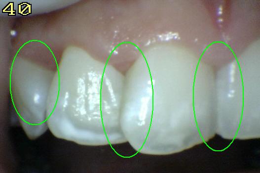
Sound tooth surface: Code 0
There should be no evidence of caries (either no or questionable
change in enamel translucency after prolonged air drying (suggested
drying time 5 seconds)). Surfaces with developmental defects
such as enamel hypoplasias; fluorosis; tooth wear (attrition,
abrasion and erosion), and extrinsic or intrinsic stains will be
recorded as sound. |
|
 |
|
|
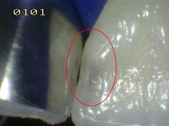
First visual change in
enamel: Code 1
When seen wet there is no evidence of any change in
color attributable to carious activity, but after prolonged air drying a
carious opacity (white or brown lesion) is visible that is not
consistent with the clinical appearance of sound enamel. This will be
seen from the buccal or lingual surface.
|
|
 |
|
|
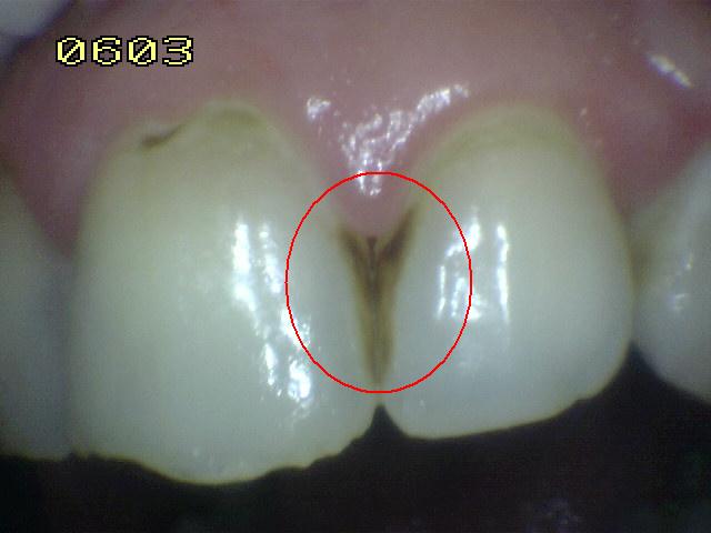
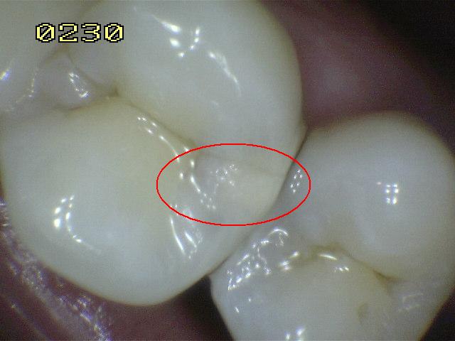
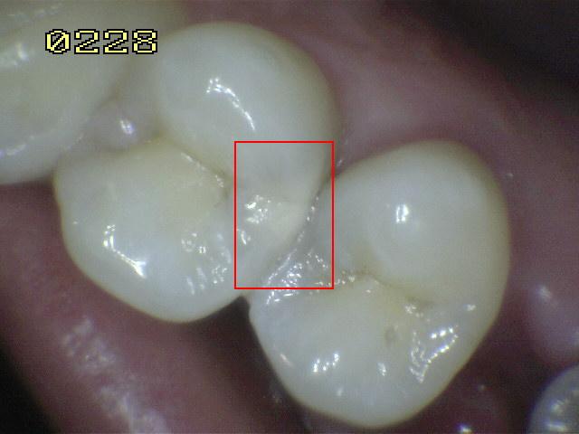
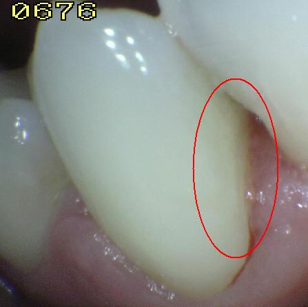
Distinct visual change in
enamel when viewed wet: Code 2
There is a carious opacity or discoloration (white or
brown lesion) that is not consistent with the clinical appearance of
sound enamel (Note: the lesion is still visible when dry). This lesion
may be seen directly when viewed from the buccal or lingual direction.
In addition, when viewed from the occlusal direction, this opacity or
discoloration may be seen as a shadow confined to enamel, seen through
the marginal ridge.
|
|
|
|
|
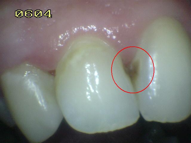
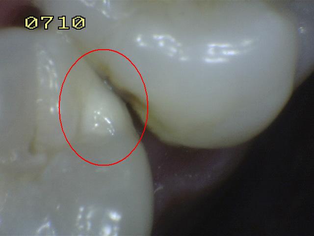
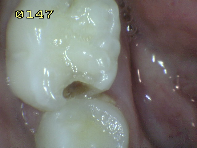
Initial breakdown in
enamel due to caries with no visible dentin: Code 3
Once dried for approximately 5 seconds there is distinct
loss of enamel integrity, viewed from the buccal or lingual direction.
If in doubt, or to confirm the visual assessment,
the
CPI probe can be used gently across the surface to confirm the loss of
surface integrity.
|
|
 |
|
Probe |
|
|
|
|
|
|
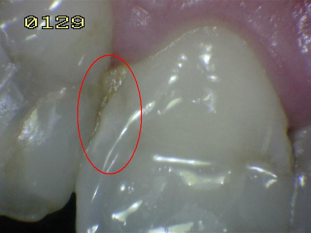
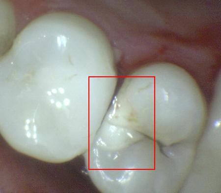
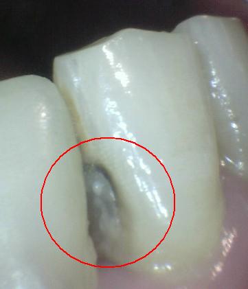
Underlying dark shadow
from dentin with or without localized enamel breakdown: Code 4
This lesion appears as a shadow of discolored dentin
visible through an apparently intact marginal ridge, buccal or lingual
walls of enamel. This appearance is often seen more easily when the
tooth is wet. The darkened area is an intrinsic shadow which may appear
as grey, blue or brown in color.
|
|
|
|
Probe |
|
|
|
|
|
|
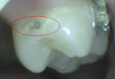
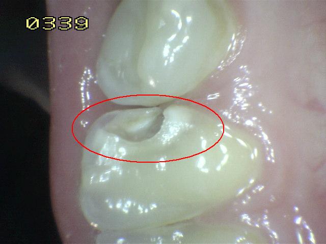
Distinct cavity with
visible dentin: Code 5.
Cavitation in opaque or discolored enamel (white or
brown) with exposed dentin in the examiner’s judgment.
If in doubt, or to confirm the visual assessment, the
CPI probe can be used to confirm the presence of a cavity apparently in
dentin. This is achieved by sliding the ball end along the surface and a
dentin cavity is detected if the ball enters the opening of the cavity
and in the opinion of the examiner the base is in dentin.
|
|
|
|
Probe |
|
|
|
|
|
|
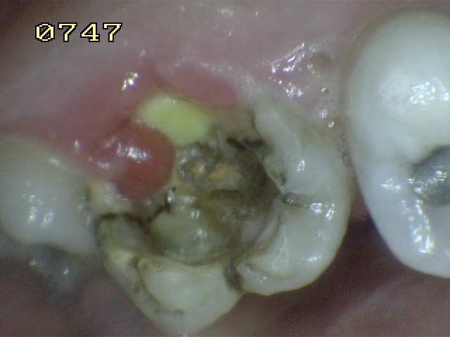
Extensive distinct cavity
with visible dentin: Code 6
Obvious loss of tooth structure, the extensive cavity
may be deep or wide and dentin is clearly visible on both the
walls and at the base. The marginal ridge may or may not be present. An
extensive cavity involves at least half of a tooth surface or possibly
reaching the pulp. |
|
 |
|
Probe |
|
|
|
 |
|